.")
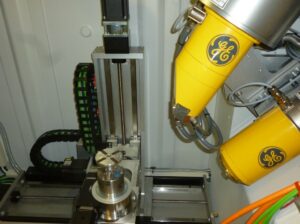
In 2015, the Regensburg Center of Biomedical Engineering (RCBE) commissioned the micro-computer tomograph (Micro-CT) at the Ostbayerische Technische Hochschule Regensburg (OTH Regensburg) funded by the German Research Foundation (DFG). The X-ray testing system phoenix v|tome|xs 240/180 from Baker Hughes Digital Solutions GmbH is interdisciplinary and is located in the Faculty of Mechanical Engineering at OTH Regensburg.
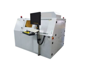 For many topics from a wide range of disciplines, it is crucial to know what it looks like “inside” an exhibit. Typically, methods that destroy or alter the exhibit are used for this purpose. Micro-CT, on the other hand, which works with X-rays, can reveal the hidden interior of objects of examination without causing damage or destruction. No special preparation, such as scanning electron microscopy, is required either. The CT also includes a high-performance graphics card-accelerated workstation with specialized software to process and visualize raw data, as well as to measure a large number of parameters.
The device is available to Campus Regensburg for research tasks and is integrated into research projects and development contracts.
The device is also available for contract research.